

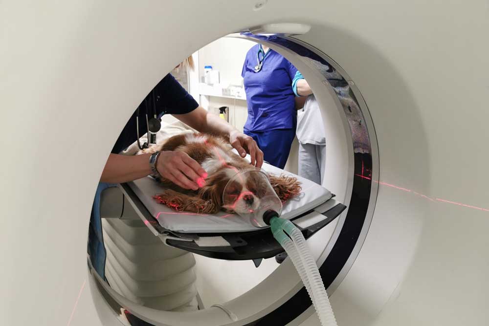
When a veterinarian recommends a CT scan after a cancer diagnosis, the goal is staging: mapping exactly where the disease sits and whether it has traveled anywhere else in the body. A physical exam and standard X-rays can only show so much, while CT builds a detailed, cross-sectional picture of the chest, abdomen, and lymph nodes where spread most often hides. For both dogs and cats, that picture frequently changes the plan, sometimes confirming that surgery is worth pursuing and sometimes sparing your pet a procedure that would not have helped. The scan is not a delay in treatment; it is how the right treatment gets chosen.
The standard we hold ourselves to at Veterinary Emergency and Specialty Center of Northern Arizona is the medicine a human hospital would deliver, and the CT scanner inside our Flagstaff facility is a big part of how we meet it. Our team performs the scan and pairs it with in-house bloodwork and chest X-rays, then explains what the images mean for the treatment paths in front of you. A staging recommendation can sound intimidating, but it exists to make the next decision clearer, not to add another hurdle. If your dog or cat has been diagnosed with cancer and a CT scan is on the table, reach out and we will answer your questions about the scan.
The Quick Map of Cancer Staging
- Staging tells us two things after a diagnosis: how much cancer there is, and whether it has spread beyond where it started.
- Metastasis means cancer cells have broken away from the original tumor and started new growths elsewhere, most often in the lymph nodes, lungs, liver, or spleen.
- Because a CT scan views the body in thin cross-sectional slices, it can catch small lesions and hidden spread that flat X-rays and ultrasound routinely miss.
- Whether cancer has spread is the single biggest factor in choosing treatment: it determines whether removing the tumor can truly help, or whether a whole-body plan makes more sense.
What Does Cancer Staging Actually Mean?
Cancer staging is the process of finding out how much disease is present and whether it has traveled beyond the spot where it first appeared. It’s a standard, expected step after a diagnosis, not a red flag, and it gives us the information we need to choose a treatment that fits your pet.
Two terms get mixed up constantly in the exam room. Grading and staging sound interchangeable, but they answer different questions about the same tumor.
| Grading | Staging | |
| What it looks at | How aggressive the tumor cells appear under a microscope | How far the cancer has spread through the body |
| How it’s done | Examining a tissue or cell sample | Imaging, bloodwork, and sampling of lymph nodes and organs |
| What it tells us | How the tumor is likely to behave | Where the disease is and which treatment fits |
What Does a Full Staging Workup Involve?
A complete staging workup reaches beyond imaging to include a physical exam, bloodwork, a sample of the tumor itself, and a look at the lymph nodes where cancer tends to settle first. Which tests belong in a workup isn’t guesswork, and current oncology guidelines match the diagnostics to the specific tumor type.
Our team can collect cytology samples, run the labwork, and read it all alongside the scan, so the pieces are interpreted together instead of one at a time.
What Is Metastasis, and Why Is Finding It So Important?
Metastasis is the process of cancer cells breaking away from the original tumor, traveling through the body, and starting new growths somewhere else. It is the single most important thing staging is designed to find, because a cancer that has spread is no longer a local problem that surgery alone can solve. It has become a whole-body disease.
Here’s how it happens. As a tumor grows, some of its cells can slip into the bloodstream or the lymphatic system, the body’s network of drainage vessels and small filtering stations called lymph nodes. Those traveling cells ride along until they lodge somewhere new, most often the lymph nodes closest to the tumor, the lungs, the liver, or the spleen, and begin building a colony of their own. A spread tumor keeps its original identity: osteosarcoma that lands in the lungs is still osteosarcoma, and it behaves with the same aggression it showed in the bone.
The lymph nodes matter so much in staging because they are usually the first stop. Cancer that is on the move tends to show up in the nearest node before it appears anywhere else, which is why we examine, image, and often sample those nodes even when they feel normal. And that word “feel” is the catch: metastatic deposits are frequently silent. A pet can have measurable spread in the lungs or a lymph node and still be eating well, playing, and acting entirely like themselves. Waiting for symptoms to reveal spread means finding it late. Imaging is how we find it early, while there are still more options on the table.
Why Does Spread Change the Entire Treatment Conversation?
Think of it as the difference between a weed and scattered seeds. If the weed hasn’t gone to seed, pulling it out solves the problem completely. Once seeds have scattered, pulling the original weed still might be worthwhile, but it no longer clears the garden. A tumor that is confined to one place can potentially be cured by removing it or treating it with focused radiation. Once cells have seeded other organs, taking out the original lump no longer removes the disease, and the plan has to account for cancer we can’t see as well as the cancer we can.
Why Is CT Such a Valuable Staging Tool?
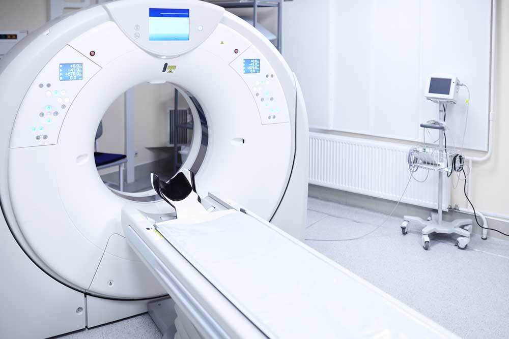
A CT scan earns its place in staging because cancer imaging technology captures the body in thin cross-sectional slices and rebuilds them into a three-dimensional picture, revealing detail that a flat X-ray or an ultrasound can’t.
Picture a standard X-ray as a single flat photograph, with everything in the chest layered on top of everything else. A CT scan takes that same region and separates it into hundreds of thin slices, so a small nodule tucked behind the heart or a mass wrapping around a blood vessel stops hiding. That resolution lets us measure a tumor precisely, find spread while it’s still tiny, and see how a growth relates to the structures around it.
| Tool | What it shows | Best for |
| X-ray | Flat, overlapping shadows of bone and soft tissue | A fast first look at the chest, belly, or a limb |
| Ultrasound | Real-time images of soft tissue and organs | Examining the abdomen and guiding samples |
| CT scan | Cross-sectional slices rebuilt in 3D | Mapping a tumor’s borders and finding small or hidden spread |
These tools answer different questions, and when a diagnosis calls for a precise map, a CT scan is the one we turn to.
What Happens During Your Pet’s CT Scan?
The appointment is shorter and calmer than most families expect. The CT scan itself takes only minutes, though most pets are sedated or briefly anesthetized so they hold completely still, and a contrast dye is often given to sharpen the images.
Even a small twitch blurs the slices, so the body has to stay quiet for those few minutes. The contrast, delivered through a small IV catheter, makes blood vessels and abnormal tissue stand out so nothing important blends into the background. Many pets are ready to head home once they’re fully awake and steady on their feet. Results are typically back within 1 to 3 days, and we’ll either go over the results with you or send the report to your regular veterinarian so that you can create a plan together.
Which Cancers Are Commonly Staged With a CT Scan?
A CT scan isn’t part of the plan for every tumor. It proves most useful when a tumor sits somewhere hard to evaluate or when knowing the exact extent of disease will change what treatment can be offered.
Tumors of the Nose, Chest, and Lungs
Nasal tumors hide within a maze of thin bone and air pockets that ordinary X-rays can’t untangle, and CT is the tool that sorts that maze into readable slices. Because the nasal cavity sits so close to the brain, a growing mass there can trigger neurologic signs alongside sneezing and nasal discharge, and a scan shows how far it has reached.
The chest is the other classic example. The lungs are one of the most common landing spots for metastasis, which is why chest imaging belongs in nearly every staging plan. Lung cancer in cats and dogs can also start in the chest itself, seeding nodules too small to catch on a chest X-ray. A tumor pressing on the airways can leave your dog or cat with labored breathing, which is worth an urgent look.
Bone, Soft Tissue, and Vascular Tumors
For tumors in bone, muscle, or the walls of blood vessels, staging is often what makes surgery possible to plan. Osteosarcoma, the most common bone cancer in dogs, is aggressive and prone to spreading, so mapping how much bone is involved and whether it has reached the lungs directly shapes what surgery can offer. Because it weakens bone from the inside, the first sign is sometimes an unexpected fracture from an ordinary jump or a misstep on a Flagstaff trail.
A soft tissue sarcoma often sends roots deeper into the surrounding tissue than its surface suggests, so knowing its true borders is what lets a surgeon plan to remove all of it the first time. Hemangiosarcoma grows from the cells lining blood vessels and often takes hold quietly in organs like the spleen and heart, where a rupture can cause sudden internal bleeding. Finding every hidden site before any plan is set is a priority with this cancer, because it spreads early and often.
How Does Staging Shape Treatment Decisions?
Staging is what turns a diagnosis into a plan. Once we know where the disease sits, the realistic options come into focus: whether surgery makes sense and how it would be approached, whether other therapies fit, and how each choice measures up against your pet’s quality of life. Deciding how to treat your pet’s cancer can be a difficult decision.
The clearest way to see it is through the fork in the road that spread creates:
- No spread found: the cancer is a local problem, and local treatments carry real weight. Surgery may remove the disease entirely, and focused options like radiation therapy become possible to aim, since a beam can only be targeted once the exact size and location of a tumor are known.
- Spread found: the goal shifts from removing the cancer to controlling it. Surgery may still have a role, easing pain or stopping a bleeding mass, but the plan now has to address disease throughout the body, which is where whole-body treatments like chemotherapy, or a comfort-focused approach, enter the conversation.
Knowing which path you’re on protects your family from two hard mistakes: pursuing an aggressive treatment that was never going to help, and turning down one that could have. Without staging, those decisions are guesses. With it, they’re informed choices.
We don’t make any of these calls for you. Our team lays out what the scan and the bloodwork reveal, walks through the paths in front of you, and talks through where each treatment can be performed, so the next decision is yours to make with a clear head.
How Does Imaging Track Progress Over Time?
That first scan does more than guide the opening move: it sets a baseline. Every scan that follows gets compared against it, which is how we tell whether a treatment is shrinking a tumor, holding it steady, or losing ground. Catching a change early means the plan can shift while there’s still room to shift it.
What If Staging Brings Hard News?
Sometimes the scan finds more disease than anyone hoped. That’s a hard day, and there’s no way around it. Even then, the information carries real value: it lets a family choose comfort on purpose rather than by default. Knowing what you’re facing means you can focus the time you have on what matters most to your pet, whether that’s pain control, a gentler pace, or simply more good days at home. We stay in that conversation with you.
Frequently Asked Questions About CT Staging for Pets
Is a CT scan safe if my pet has cancer?
A CT scan is safe for the large majority of pets, and a cancer diagnosis rarely changes that. The scan itself is painless and quick. Any real risk comes from the sedation or brief anesthesia that keeps your pet still, which is why we check bloodwork and weigh your pet’s age and overall health first. Dogs and cats with heart or kidney concerns get extra care, especially around the contrast dye.
Why can’t we just remove the tumor and skip the staging?
Because surgery only helps if the tumor is the whole problem. If cancer has already seeded the lungs or lymph nodes, removing the original mass puts your pet through anesthesia and recovery without changing the course of the disease. Staging first tells us whether surgery can actually deliver what you’re hoping for, and if it can’t, it points to the options that still can help.
What does it mean if staging shows my pet’s cancer has spread?
It means cancer cells have traveled from the original tumor and established new growths elsewhere, so the disease is now a whole-body condition rather than a single lump. That changes the goal of treatment, but it does not automatically mean nothing can be done. Some pets with metastatic cancer enjoy meaningful, comfortable time with the right plan, and every pet’s situation is different.
What does a CT scan cost?
The cost of a CT scan varies depending on what body parts need scanning, the exact type of scan we’re doing, the size of your pet, whether we’re sampling any masses or lymph nodes, and how long they need to be anesthetized. We’ll provide you a clear estimate of the costs, and we provide CareCredit and ScratchPay to help spread the costs over time.
Moving Forward With Clear Information
A recommendation to stage your pet’s cancer isn’t a stall or a bad omen. It’s the sign of a team that would rather build a plan around the whole picture than guess at it. Whatever the scan shows, you won’t be left to sort through it alone. When a CT scan is part of your pet’s cancer plan, reach us at Veterinary Emergency and Specialty Center of Northern Arizona in Flagstaff and we’ll walk you through what the images mean and what comes next.





Leave A Comment